
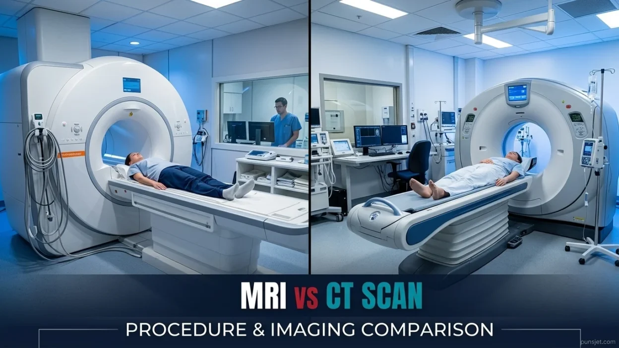
Getting the right diagnosis often starts with choosing the right imaging test. When a doctor recommends an MRI scan or a CT scan, you may wonder how they differ and why one is recommended over the other. Both tests help doctors examine the inside of the body without surgery, but they use different technologies and offer different advantages.
The main difference between MRI and CT scans is how they create images. MRI (magnetic resonance imaging) uses strong magnets and radio waves to produce detailed images of soft tissues, while CT (computed tomography) uses X-rays to create detailed cross-sectional images of the body. MRI is often useful for examining the brain, spinal cord, muscles, and ligaments. CT scans are generally faster and commonly used for injuries, internal bleeding, lung conditions, and other urgent medical problems. The right test depends on your symptoms, medical history, and the information your doctor needs.
Quick Answer: MRI vs CT Scan
An MRI uses magnets and radio waves, while a CT scan uses X-rays. MRI often provides better detail of soft tissues, including the brain, spinal cord, and ligaments. CT scans are generally faster and useful for emergencies, fractures, and internal bleeding. Neither test is always better; the best choice depends on the medical condition.
Why Do People Confuse MRI and CT Scans?
People often confuse MRI and CT scans because both produce detailed images of the inside of the body. Doctors use them to investigate symptoms, identify diseases, and plan treatments.
Both tests can examine the brain, chest, abdomen, and spine. However, their technologies, scanning times, and strengths are different.
Here are the main differences:
- MRI uses strong magnets and radio waves.
- CT scan uses X-rays and computer processing.
- Soft-tissue imaging is one of MRI’s major strengths.
- Fast imaging is one of CT’s main advantages.
- Ionizing radiation is used in CT scans but not in MRI scans.
- Contrast material may be used with either test to make certain structures easier to see.
Doctors consider these differences when choosing the most appropriate examination for a patient.
First Word: What Is an MRI Scan?
MRI stands for magnetic resonance imaging. It is a medical imaging test that uses a strong magnetic field, radio waves, and a computer to create detailed pictures of structures inside the body.
Unlike a CT scan, an MRI does not use X-rays or ionizing radiation. This can be an advantage for patients who need repeated imaging, although MRI still has specific safety requirements.
What Does an MRI Scan Show?
MRI is particularly useful for examining soft tissues, including muscles, ligaments, tendons, nerves, the brain, and the spinal cord.
Doctors may recommend an MRI to investigate:
- Brain abnormalities and certain brain tumors.
- Spinal cord and nerve problems.
- Torn ligaments and tendon injuries.
- Muscle and joint conditions.
- Certain causes of back pain.
- Some liver and pelvic conditions.
- Certain cancers, including selected prostate, breast, and liver cancers.
- Possible cancer spread to the brain or bones.
MRI can reveal subtle changes in some tissues that may be difficult to see clearly with other imaging methods. However, the best test depends on the condition being investigated.
How Does an MRI Work?
During an MRI, you lie on a movable table that slides into a cylindrical scanner. The machine uses a magnetic field and radio waves to generate signals from the body’s tissues. A computer processes these signals into detailed images.
The scanner makes loud tapping or thumping sounds during the examination. Patients are usually provided with earplugs or headphones to reduce the noise.
An MRI examination commonly takes around 20–60 minutes, although some examinations are shorter or longer. You must remain still to help the machine produce clear images.
Because the scanner can feel enclosed, some people experience claustrophobia, which means fear or discomfort in tight or enclosed spaces. If you are worried about this, tell your healthcare team before the examination. They may discuss ways to help you feel more comfortable.
What Are the Risks of an MRI?
MRI does not expose patients to ionizing radiation, but its powerful magnet requires careful safety screening.
Before the examination, tell the medical team if you have a pacemaker, an implanted medical device, metal fragments, or a history of metal-related injuries. Some newer devices are MRI-compatible under specific conditions, so the team must verify their safety rather than assume all implants are unsuitable.
Some MRI examinations use a gadolinium-based contrast agent to make certain tissues and abnormalities easier to see. The medical team considers factors such as kidney function, previous reactions, and pregnancy when deciding whether contrast is appropriate.
MRI is generally well tolerated, but the examination may take longer than CT and can be uncomfortable for people who find enclosed spaces difficult.
Second Word: What Is a CT Scan?
CT stands for computed tomography. A CT scan uses X-rays and computer technology to create detailed cross-sectional images of the body.
These images show different layers of the body, helping doctors examine bones, organs, blood vessels, and other structures. A CT scan is also sometimes called a CAT scan.
What Does a CT Scan Show?
CT scans are useful for examining many body parts, particularly when doctors need information quickly.
Doctors may recommend a CT scan to investigate:
- Broken bones and serious injuries.
- Internal bleeding after an accident.
- Certain head injuries and possible strokes.
- Lung and chest conditions.
- Blood clots in selected clinical situations.
- Kidney stones.
- Bowel blockage.
- Certain tumors and infections.
- Injuries to internal organs.
- Possible cancer spread to organs such as the lungs and liver.
CT scans provide a broad view of internal structures in a short period. This makes them particularly useful when doctors need to make quick decisions about diagnosis or treatment.
How Does a CT Scan Work?
During a CT scan, you lie on a table that moves through a large, ring-shaped machine. An X-ray source rotates around your body and takes images from different angles.
A computer combines the information to create detailed cross-sectional pictures. These images help doctors examine internal structures more closely than a single standard X-ray image would allow.
The image acquisition often takes only a few minutes, although preparation and the full appointment may take longer. You may be asked to hold your breath briefly to help produce clear images.
Some CT examinations use iodine-based contrast material to make certain organs, blood vessels, or abnormalities easier to see. The contrast may be injected into a vein or, for certain examinations, swallowed.
What Are the Risks of a CT Scan?
CT scans use ionizing radiation. The risk from an individual examination is generally small, but exposure can contribute to a person’s lifetime risk of cancer. The level of risk depends on factors such as the radiation dose, age, and type of examination.
Doctors recommend CT scans when the expected medical benefits outweigh the potential risks. They also use appropriate scanning techniques to limit unnecessary radiation exposure.
CT contrast material can cause reactions in some patients. Kidney function may also need consideration before certain contrast-enhanced examinations.
Tell your healthcare team if you are pregnant or might be pregnant, have kidney problems, or have previously experienced a reaction to contrast material.
MRI vs CT Scan: Comparison Table
| Feature | MRI Scan | CT Scan |
|---|---|---|
| Full name | Magnetic resonance imaging | Computed tomography |
| Technology | Strong magnets and radio waves | X-rays and computer processing |
| Ionizing radiation | No | Yes |
| Soft-tissue detail | Often excellent | Good, depending on the condition |
| Bone imaging | Useful for bone marrow and surrounding tissues | Often excellent for fractures |
| Common uses | Brain, spinal cord, ligaments, muscles | Injuries, bleeding, lungs, bones |
| Typical scan time | Often 20–60 minutes | Often a few minutes for image acquisition |
| Noise | Usually loud | Usually quieter |
| Enclosed space | Cylindrical scanner may cause discomfort | Usually a wider, ring-shaped scanner |
| Metal implants | Requires careful safety screening | Usually less affected by implants |
| Contrast material | May use gadolinium | May use iodine-based contrast |
| Emergency use | Useful in selected situations | Often preferred when rapid imaging is needed |
| Cancer assessment | Useful for detailed views of selected tissues and organs | Useful for assessing organs and possible cancer spread |
These are general comparisons. The most appropriate examination depends on the medical condition, the body part being examined, and how quickly the information is needed.
When Should You Use an MRI or a CT Scan?
Doctors choose MRI or CT based on the medical question they need to answer. Sometimes a CT scan is performed first, followed by an MRI if more detailed information is needed.
When Is an MRI Scan Better?
An MRI is often preferred when detailed images of soft tissues are important.
Examples include:
- Brain conditions: MRI can help investigate certain tumors and other brain abnormalities.
- Spinal problems: It can show spinal discs, nerves, and the spinal cord.
- Sports injuries: MRI can help identify torn ligaments, tendons, and muscles.
- Joint problems: It can show damage to structures inside the knee, shoulder, and other joints.
- Prostate and breast conditions: MRI may provide detailed information for selected diagnostic questions.
- Liver conditions: MRI can help characterize certain liver abnormalities.
- Possible cancer spread: MRI can provide detailed information about selected abnormalities in the brain and bones.
MRI is not always the first choice for these conditions. A CT scan may be more appropriate when doctors need to identify fractures or urgent problems quickly.
When Is a CT Scan Better?
A CT scan is often preferred when speed is important or doctors need detailed images of bones, lungs, or internal injuries.
Common examples include:
- Serious accidents: CT can help identify internal injuries quickly.
- Possible internal bleeding: It can help doctors locate bleeding after trauma.
- Head injuries: CT is commonly used to assess bleeding and skull fractures.
- Lung conditions: CT provides detailed images of the lungs and chest.
- Cancer assessment: CT can help doctors evaluate whether cancer may have spread to other organs.
- Kidney stones: CT can help detect stones and determine their size and location.
- Bowel blockage: CT can help identify an obstruction in the intestines.
- Complex fractures: It can show the position and shape of broken bones.
CT scans are especially valuable in emergencies because they can provide important information quickly and help doctors decide on immediate treatment.
Which Is Better for a Brain Scan?
Neither MRI nor CT is always better for examining the brain.
CT is commonly used in emergencies when doctors need to check for bleeding, skull fractures, or certain signs of stroke. MRI can provide more detailed information about some brain conditions, including certain tumors and small abnormalities.
If a doctor suspects that cancer has spread to the brain, MRI may be recommended because it can reveal subtle changes in brain tissue. The correct test depends on the symptoms, suspected condition, and urgency.
Which Is Better for Back Pain?
MRI is often more useful when doctors suspect a problem involving spinal discs, nerves, or the spinal cord.
CT can be helpful for certain spinal fractures and may be used when MRI is unsuitable. However, many cases of back pain do not require immediate imaging.
A doctor will consider the symptoms, medical history, and physical examination before recommending a scan.
Which Is Safer: MRI or CT?
MRI avoids ionizing radiation, which can be an advantage when radiation exposure is a concern. However, MRI requires careful screening for metal objects and certain implanted devices.
CT uses radiation but can provide essential information quickly. When CT is medically necessary, avoiding it solely because of radiation concerns could delay diagnosis or treatment.
The safest option is the examination that provides the required information while appropriately managing the risks for the individual patient.
What Concerns Do People Have About MRI and CT Scans?
Both MRI and CT scans are noninvasive imaging tests, meaning they allow doctors to examine internal structures without performing surgery. Most patients tolerate these examinations well, but each test has different considerations.
Concerns About CT Scans
The main concern about CT is radiation exposure. The radiation dose is controlled, and doctors consider whether the expected benefits justify the examination.
Some CT scans also use contrast material. Most people tolerate it, but allergic-like reactions and other complications can occur. The medical team reviews relevant medical history before administering contrast when needed.
CT scanners generally have a ring-shaped opening and are usually easier for people who dislike enclosed spaces. CT may also be suitable for many patients with medical implants that require special precautions during MRI.
Concerns About MRI Scans
MRI does not use ionizing radiation, but the examination takes longer and the scanner can feel enclosed. People with claustrophobia may find it uncomfortable.
The machine also makes loud noises, so ear protection is normally provided. Patients must remain still to help produce clear images.
Some pacemakers, stimulators, and other implants may restrict MRI use. However, many newer devices can be scanned under specific safety conditions.
Like CT, MRI may involve contrast material, depending on the medical question. The medical team determines whether it is necessary and considers any relevant risks.
If you have concerns about radiation, contrast, enclosed spaces, or implanted devices, discuss them with your healthcare team before the examination.
How Is Artificial Intelligence Improving MRI and CT Scan Imaging?
Artificial intelligence (AI) is becoming a useful tool in medical imaging. It can assist radiologists by identifying patterns in images, improving image quality, and helping them examine particular areas more closely.
For example, AI-assisted tools may help radiologists assess changes in a tumor over time, including whether it appears to have grown or shrunk. These tools can also help highlight areas that may need further review.
However, AI does not replace radiologists. A trained medical professional must interpret the images alongside the patient’s symptoms, medical history, and other relevant findings. AI is an additional tool that may support the review process, not a substitute for professional medical judgment.
The availability and use of AI tools vary between healthcare facilities, and not every MRI or CT examination uses them.
How Do Doctors Choose the Right Scan for You?
Doctors personalize imaging decisions based on your symptoms, medical history, physical examination, and suspected condition.
In many situations, CT is selected first because it provides a quick, broad view of the body. If the scan reveals something that needs closer examination, an MRI may be recommended for more detailed information.
For example, a CT scan might identify an abnormal area in an organ, while a follow-up MRI may help doctors characterize that area more clearly. In other cases, MRI may be the preferred first examination.
Doctors also consider practical factors, including:
- The suspected condition: Which test is most likely to answer the medical question?
- Urgency: Does the patient need rapid imaging?
- Radiation exposure: Can the condition be assessed effectively without ionizing radiation?
- Medical implants: Is MRI safe for the patient’s particular device?
- Comfort: Does the patient have claustrophobia or difficulty remaining still?
- Contrast material: Is contrast necessary, and are there relevant risks?
- Previous imaging: Have earlier examinations already provided useful information?
The goal is to obtain the clearest useful images while considering safety, comfort, and the patient’s individual needs.
12 Examples of MRI vs CT Scan in Medical Situations
These examples show why doctors may choose one scan over the other.
- A serious head injury: A CT scan may be used to check quickly for bleeding or a skull fracture.
- Ongoing knee pain: An MRI may help identify damage to ligaments, cartilage, or tendons.
- Sudden weakness on one side of the body: Emergency imaging may begin with CT, depending on the suspected cause and local stroke protocol.
- A suspected spinal disc problem: MRI can provide detailed images of the discs and nearby nerves.
- A car accident: CT may help doctors assess internal injuries rapidly.
- A suspected brain tumor: MRI often provides detailed information about the tumor and surrounding brain tissue.
- Possible kidney stones: CT can help identify stones in the urinary tract.
- A complex fracture: CT can show the shape and position of broken bone fragments.
- A possible spinal cord injury: MRI may help assess damage to the spinal cord and surrounding soft tissues.
- A lung condition: CT can provide detailed images of the lungs and chest.
- A suspected torn shoulder tendon: MRI may show the location and extent of the injury.
- Unexplained abdominal pain: CT or MRI may be recommended depending on the symptoms and suspected cause.
These examples are general guides. A doctor decides which examination is most suitable for each patient.
Common Mistakes About MRI and CT Scans
People sometimes misunderstand the differences between these medical imaging tests.
Mistake 1: Thinking MRI and CT Use the Same Technology
MRI uses magnets and radio waves, while CT uses X-rays. Both produce images, but they work differently.
Mistake 2: Believing MRI Is Always Better
MRI often provides excellent soft-tissue detail, but CT is usually faster and may be more suitable for certain injuries and emergencies.
Mistake 3: Assuming CT Scans Are Always Dangerous
CT uses ionizing radiation, but the risks of a medically necessary examination are generally small compared with its potential benefits. Doctors consider these risks before recommending the test.
Mistake 4: Believing MRI Has No Safety Risks
MRI does not use ionizing radiation, but its strong magnetic field creates safety concerns for certain implants and metal objects.
Mistake 5: Thinking Every Scan Requires Contrast
Many MRI and CT examinations are performed without contrast. Whether contrast material is needed depends on the medical question.
Mistake 6: Assuming Every Medical Problem Requires Imaging
Some conditions can be diagnosed through symptoms, medical history, and a physical examination. Imaging is recommended when it is likely to provide useful information.
Mistake 7: Thinking Artificial Intelligence Makes the Final Diagnosis
Artificial intelligence may help analyze images, but radiologists interpret the findings and consider the patient’s medical information before reaching a conclusion.
Memory Trick: How to Remember MRI vs CT Scan
A simple memory trick can help you remember the difference.
- MRI = Magnets: Think of magnets and detailed images of soft tissues.
- CT = Quick Test: Think of fast X-ray imaging that is useful in many emergencies.
Remember that CT is not always the fastest option for every situation, and MRI can also be performed quickly in certain circumstances. This trick is simply a way to remember their main differences.
American vs British English
MRI and CT scan are standard medical terms in both American and British English.
| Term | American English | British English |
|---|---|---|
| MRI | MRI scan | MRI scan |
| CT | CT scan or CAT scan | CT scan |
| MRI full name | Magnetic resonance imaging | Magnetic resonance imaging |
| CT full name | Computed tomography | Computed tomography |
| Medical specialist | Radiologist | Radiologist |
There is no significant spelling difference between these terms in American and British English. Both varieties use them to describe medical imaging tests.
Similar Words and Medical Imaging Terms
Several related medical terms can help you understand MRI and CT scans more clearly.
- X-ray: Uses radiation to create images, often of bones and the chest. CT scans use multiple X-ray measurements to produce cross-sectional images.
- Ultrasound: Uses sound waves to create images. It is commonly used during pregnancy and to examine certain organs and blood vessels.
- PET scan: Uses a radioactive tracer to show information about activity in body tissues. It may be combined with CT or MRI.
- Contrast agent: A substance used during certain scans to make particular structures easier to see.
- Radiologist: A medical doctor trained to interpret medical images and help guide imaging decisions.
- Radiation exposure: The amount of ionizing radiation a person receives during an examination such as a CT scan.
- Claustrophobia: Fear or discomfort associated with enclosed spaces, which can make some MRI examinations challenging.
- Cross-sectional imaging: A method of showing the body in slices, allowing doctors to examine internal structures layer by layer.
- Computed tomography: The full name of CT, which uses X-rays and computer processing to create detailed images.
Knowing these terms can make it easier to understand your doctor’s explanation of the available imaging options.
FAQs:
1. What Is the Main Difference Between an MRI and a CT Scan?
The main difference is the technology used. MRI uses strong magnets and radio waves, while CT uses X-rays. MRI often provides better soft-tissue detail, whereas CT is generally faster and useful for many injuries and emergencies.
2. Is an MRI Better Than a CT Scan?
Not always. MRI is often better for detailed images of soft tissues, nerves, and ligaments. CT is often better for quickly identifying certain fractures, internal bleeding, and lung problems. The best test depends on the medical condition.
3. Which Scan Is Faster, MRI or CT?
CT scans are generally faster. Image acquisition often takes only a few minutes, while MRI examinations commonly take 20–60 minutes. The actual time varies depending on the body part and examination.
4. Does an MRI Have Radiation?
No. MRI uses magnetic fields and radio waves rather than ionizing radiation. However, patients must follow MRI safety procedures, particularly if they have medical implants or metal in their bodies.
5. Is a CT Scan Safe?
CT scans are generally considered appropriate when medically necessary. They use ionizing radiation, which carries a small potential long-term risk. Doctors weigh this risk against the benefits of obtaining information needed for diagnosis or treatment.
6. Which Is Better for Brain Problems, MRI or CT?
It depends on the suspected condition. CT is commonly used for urgent assessment of head injuries and possible bleeding. MRI can provide more detailed information about certain brain conditions, including some tumors and small abnormalities.
7. Can an MRI Detect Problems That a CT Scan Misses?
Yes, in some situations. MRI can reveal certain soft-tissue abnormalities that are difficult to see on CT. However, CT can identify problems that MRI may not show as clearly or as quickly. Neither test detects every condition.
8. Do MRI and CT Scans Require Contrast Dye?
No. Many scans can be performed without contrast. Some MRI examinations use gadolinium-based contrast, while some CT examinations use iodine-based contrast. The medical team decides whether contrast is needed and considers any relevant risks.
Remember This
- MRI uses magnets and radio waves.
- CT scans use X-rays.
- MRI often provides better soft-tissue detail.
- CT is generally faster and useful in many emergencies.
- MRI does not use ionizing radiation, but it has specific safety requirements.
- CT uses radiation, so unnecessary examinations should be avoided.
- Contrast material may be used with either test.
- Artificial intelligence can assist image analysis but does not replace a radiologist’s medical judgment.
- The right scan depends on the medical problem, not simply on which technology is more advanced.
Conclusion
The difference between MRI and CT scans comes down to how they create images, what they show best, and how quickly doctors need the results. MRI is particularly useful for examining soft tissues, including the brain, spinal cord, ligaments, and muscles. CT is especially useful for rapidly assessing injuries, fractures, internal bleeding, and many chest or abdominal problems. Both tests help doctors make informed decisions about diagnosis and treatment.
Doctors may also consider radiation exposure, contrast material, medical implants, and patient comfort when selecting a scan. Some patients benefit from a CT scan first, followed by an MRI for a closer examination. Others may need only one test. If your doctor recommends either examination, ask why it is appropriate for your condition and share any relevant medical concerns. Understanding these differences can help you feel more prepared and make informed decisions with your healthcare team.

Donald Andrew is the creative mind behind PunsJet.com, where he shares clever wordplay and humorous puns for readers around the world. With a passion for language and comedy, Donald enjoys crafting witty jokes that bring smiles to everyday conversations. His goal is to make humor simple, fun, and easy to share.